For a better understanding of tissue organisation of roots, stems and leaves, it is convenient to study the transverse sections of the mature zones of these organs.
Look at Figure 6.6 (a), it shows the transverse section of the sunflower root. The internal tissue organisation is as follows:
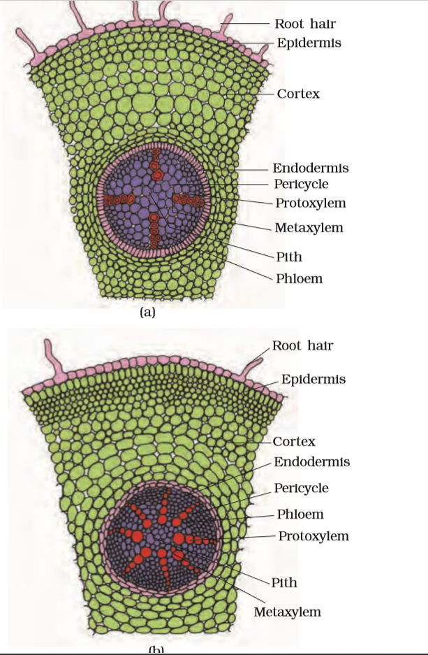
Figure 6.6 T.S. : (a) Dicot root (Primary) (b) Monocot root
The outermost layer is epiblema. Many of the cells of epiblema protrude in the form of unicellular root hairs. The cortex consists of several layers of thin-walled parenchyma cells with intercellular spaces. The innermost layer of the cortex is called endodermis. It comprises a single layer of barrel-shaped cells without any intercellular spaces. The tangential as well as radial walls of the endodermal cells have a deposition of water-impermeable, waxy material suberin in the form of casparian strips. Next to endodermis lies a few layers of thick-walled parenchyomatous cells referred to as pericycle. Initiation of lateral roots and vascular cambium during the secondary growth takes place in these cells. The pith is small or inconspicuous. The parenchymatous cells which lie between the xylem and the phloem are called conjuctive tissue. There are usually two to four xylem and phloem patches. Later, a cambium ring develops between the xylem and phloem. All tissues on the innerside of the endodermis such as pericycle, vascular bundles and pith constitute the stele.
The anatomy of the monocot root is similar to the dicot root in many respects (Figure 6.6 b). It has epidermis, cortex, endodermis, pericycle, vascular bundles and pith. As compared to the dicot root which have fewer xylem bundles, there are usually more than six (polyarch) xylem bundles in the monocot root. Pith is large and well developed. Monocotyledonous roots do not undergo any secondary growth.

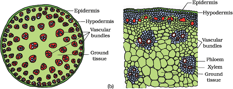
Figure 6.7 T.S. of stem : (a) Dicot (b) Monocot
© 2026 GoodEd Technologies Pvt. Ltd.

