The alimentary canal present in the body cavity is divided into three regions: foregut, midgut and hindgut (Figure 7.16). The mouth opens into a short tubular pharynx, leading to a narrow tubular passage called oesophagus. This in turn opens into a sac like structure called crop used for storing of food. The crop is followed by gizzard or proventriculus. It has an outer layer of thick circular muscles and thick inner cuticle forming six highly chitinous plate called teeth. Gizzard helps in grinding the food particles. The entire foregut is lined by cuticle. A ring of 6-8 blind tubules called hepatic or gastric caeca is present at the junction of foregut and midgut, which secrete digestive juice. At the junction of midgut and hindgut is present another ring of 100-150 yellow coloured thin filamentous Malpighian tubules. They help in removal of excretory products from haemolymph. The hindgut is broader than midgut and is differentiated into ileum, colon and rectum. The rectum opens out through anus.
Blood vascular system of cockroach is an open type (Figure 7.17). Blood vessels are poorly developed and open into space (haemocoel). Visceral organs located in the haemocoel are bathed in blood (haemolymph). The haemolymph is composed of colourless plasma and haemocytes. Heart of cockroach consists of elongated muscular tube lying along mid dorsal line of thorax and abdomen. It is differentiated into funnel shaped chambers with ostia on either side. Blood from sinuses enter heart through ostia and is pumped anteriorly to sinuses again.
The respiratory system consists of a network of trachea, that open through 10 pairs of small holes called spiracles present on the lateral side of the body. Thin branching tubes (tracheal tubes subdivided into tracheoles) carry oxygen from the air to all the parts. The opening of the spiracles is regulated by the sphincters. Exchange of gases take place at the tracheoles by diffusion.
Excretion is performed by Malpighian tubules. Each tubule is lined by glandular and ciliated cells. They absorb nitrogenous waste products and convert them into uric acid which is excreted out through the hindgut. Therefore, this insect is called uricotelic. In addition, the fat body, nephrocytes and urecose glands also help in excretion.
The nervous system of cockroach consists of a series of fused, segmentally arranged ganglia joined by paired longitudinal connectives on the ventral side. Three ganglia lie in the thorax, and six in the abdomen. The nervous system of cockroach is spread throughout the body. The head holds a bit of a nervous system while the rest is situated along the ventral (belly-side) part of its body. So, now you understand that if the head of a cockroach is cut off, it will still live for as long as one week. In the head region, the brain is represented by supra-oesophageal ganglion which supplies nerves to antennae and compound eyes. In cockroach, the sense organs are antennae, eyes, maxillary palps, labial palps, anal cerci, etc. The compound eyes are situated at the dorsal surface of the head. Each eye consists of about 2000 hexagonal ommatidia (sing.: ommatidium). With the help of several ommatidia, a cockroach can receive several images of an object. This kind of vision is known as mosaic vision with more sensitivity but less resolution, being common during night (hence called nocturnal vision).
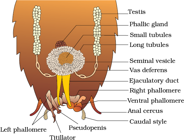
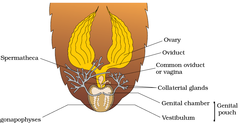
Cockroaches are dioecious and both sexes have well developed reproductive organs (Figure 7.18). Male reproductive system consists of a pair of testes one lying on each lateral side in the 4th -6th abdominal segments. From each testis arises a thin vas deferens, which opens into ejaculatory duct through seminal vesicle. The ejaculatory duct opens into male gonopore situated ventral to anus. A characteristic mushroom-shaped gland is present in the 6th-7th abdominal segments which functions as an accessory reproductive gland. The external genitalia are represented by male gonapophysis or phallomere (chitinous asymmetrical structures, surrounding the male gonopore). The sperms are stored in the seminal vesicles and are glued together in the form of bundles called spermatophores which are discharged during copulation. The female reproductive sysytem consists of two large ovaries, lying laterally in the 2nd – 6th abdominal segments. Each ovary is formed of a group of eight ovarian tubules or ovarioles, containing a chain of developing ova. Oviducts of each ovary unite into a single median oviduct (also called vagina) which opens into the genital chamber. A pair of spermatheca is present in the 6th segment which opens into the genital chamber.


Sperms are transferred through spermatophores. Their fertilised eggs are encased in capsules called oothecae. Ootheca is a dark reddish to blackish brown capsule, about 3/8” (8 mm) long. They are dropped or glued to a suitable surface, usually in a crack or crevice of high relative humidity near a food source. On an average, females produce 9-10 oothecae, each containing 14-16 eggs. The development of P. americana is paurometabolous, meaning there is development through nymphal stage. The nymphs look very much like adults. The nymph grows by moulting about 13 times to reach the adult form. The next to last nymphal stage has wing pads but only adult cockroaches have wings.
Many species of cockroaches are wild and are of no known economic importance yet. A few species thrive in and around human habitat. They are pests because they spoil food and contaminate it with their smelly excreta. They can transmit a variety of bacterial diseases by contaminating food material.