The body cavity of frogs accommodate different organ systems such as digestive, circulatory, respiratory, nervous, excretory and reproductive systems with well developed structures and functions (Figure 7.20).
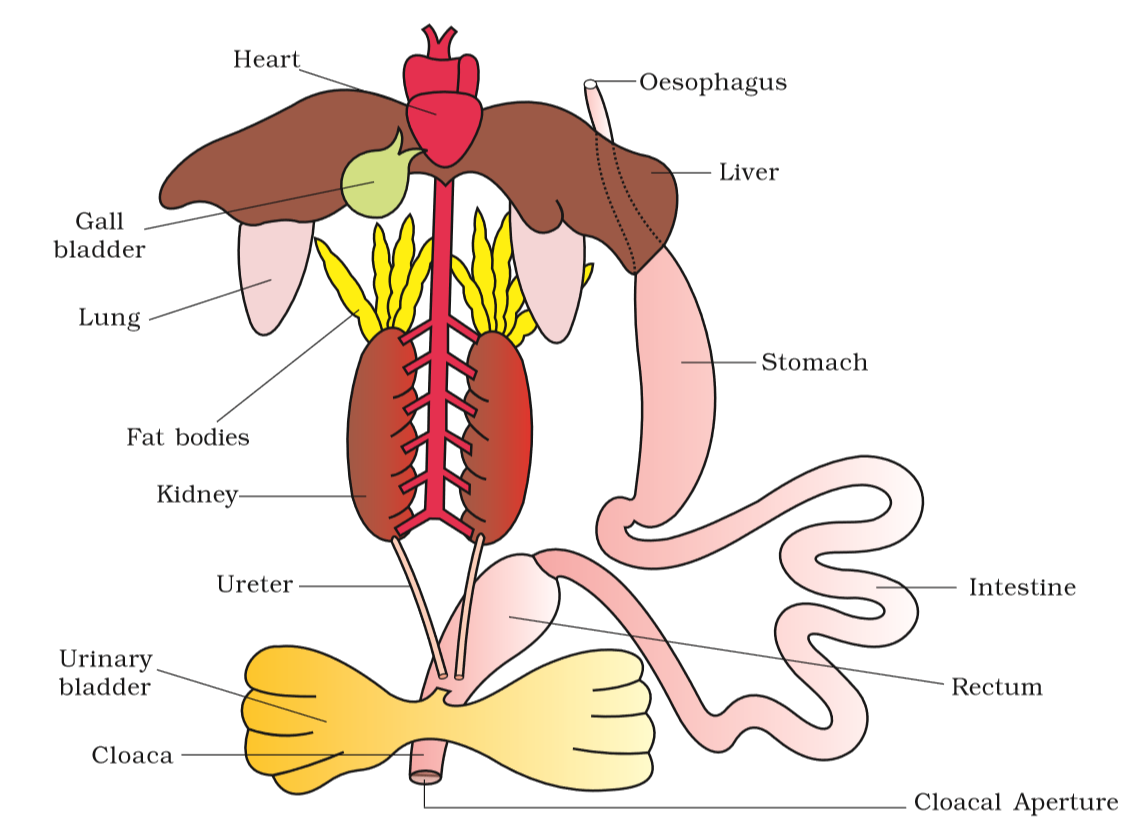
Figure 7.20 Diagrammatic representation of internal organs of frog showing complete digestive system
The digestive system consists of alimentary canal and digestive glands. The alimentary canal is short because frogs are carnivores and hence the length of intestine is reduced. The mouth opens into the buccal cavity that leads to the oesophagus through pharynx. Oesophagus is a short tube that opens into the stomach which in turn continues as the intestine, rectum and finally opens outside by the cloaca. Liver secretes bile that is stored in the gall bladder. Pancreas, a digestive gland produces pancreatic juice containing digestive enzymes. Food is captured by the bilobed tongue. Digestion of food takes place by the action of HCl and gastric juices secreted from the walls of the stomach. Partially digested food called chyme is passed from stomach to the first part of the small intestine, the duodenum. The duodenum receives bile from gall bladder and pancreatic juices from the pancreas through a common bile duct. Bile emulsifies fat and pancreatic juices digest carbohydrates and proteins. Final digestion takes place in the intestine. Digested food is absorbed by the numerous finger-like folds in the inner wall of intestine called villi and microvilli. The undigested solid waste moves into the rectum and passes out through cloaca.
Frogs respire on land and in the water by two different methods. In water, skin acts as aquatic respiratory organ (cutaneous respiration). Dissolved oxygen in the water is exchanged through the skin by diffusion. On land, the buccal cavity, skin and lungs act as the respiratory organs. The respiration by lungs is called pulmonary respiration. The lungs are a pair of elongated, pink coloured sac-like structures present in the upper part of the trunk region (thorax). Air enters through the nostrils into the buccal cavity and then to lungs. During aestivation and hibernation gaseous exchange takes place through skin.
The vascular system of frog is well-developed closed type. Frogs have a lymphatic system also. The blood vascular system involves heart, blood vessels and blood. The lymphatic system consists of lymph, lymph channels and lymph nodes. Heart is a muscular structure situated in the upper part of the body cavity. It has three chambers, two atria and one ventricle and is covered by a membrane called pericardium. A triangular structure called sinus venosus joins the right atrium. It receives blood through the major veins called vena cava. The ventricle opens into a sac-like conus arteriosus on the ventral side of the heart. The blood from the heart is carried to all parts of the body by the arteries (arterial system). The veins collect blood from different parts of body to the heart and form the venous system. Special venous connection between liver and intestine as well as the kidney and lower parts of the body are present in frogs. The former is called hepatic portal system and the latter is called renal portal system. The blood is composed of plasma and cells. The blood cells are RBC (red blood cells) or erythrocytes, WBC (white blood cells) or leucocytes and platelets. RBC’s are nucleated and contain red coloured pigment namely haemoglobin. The lymph is different from blood. It lacks few proteins and RBCs. The blood carries nutrients, gases and water to the respective sites during the circulation. The circulation of blood is achieved by the pumping action of the muscular heart.
The elimination of nitrogenous wastes is carried out by a well developed excretory system. The excretory system consists of a pair of kidneys, ureters, cloaca and urinary bladder. These are compact, dark red and bean like structures situated a little posteriorly in the body cavity on both sides of vertebral column. Each kidney is composed of several structural and functional units called uriniferous tubules or nephrons. Two ureters emerge from the kidneys in the male frogs. The ureters act as urinogenital duct which opens into the cloaca. In females the ureters and oviduct open seperately in the cloaca. The thin-walled urinary bladder is present ventral to the rectum which also opens in the cloaca. The frog excretes urea and thus is a ureotelic animal. Excretory wastes are carried by blood into the kidney where it is separated and excreted.
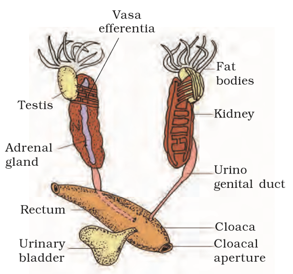
Figure 7.21 Male reproductive system
The system for control and coordination is highly evolved in the frog. It includes both neural system and endocrine glands. The chemical coordination of various organs of the body is achieved by hormones which are secreted by the endocrine glands. The prominent endocrine glands found in frog are pituitary, thyroid, parathyroid, thymus, pineal body, pancreatic islets, adrenals and gonads. The nervous system is organised into a central nervous system (brain and spinal cord), a peripheral nervous system (cranial and spinal nerves) and an autonomic nervous system (sympathetic and parasympathetic). There are ten pairs of cranial nerves arising from the brain. Brain is enclosed in a bony structure called brain box (cranium). The brain is divided into fore-brain, mid-brain and hind-brain. Forebrain includes olfactory lobes, paired cerebral hemispheres and unpaired diencephalon. The midbrain is characterised by a pair of optic lobes. Hind-brain consists of cerebellum and medulla oblongata. The medulla oblongata passes out through the foramen magnum and continues into spinal cord, which is enclosed in the vertebral column.
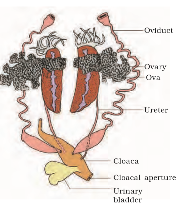
Figure 7.22 Female reproductive system
Frog has different types of sense organs, namely organs of touch (sensory papillae), taste (taste buds), smell (nasal epithelium), vision (eyes) and hearing (tympanum with internal ears). Out of these, eyes and internal ears are well-organised structures and the rest are cellular aggregations around nerve endings. Eyes in a frog are a pair of spherical structures situated in the orbit in skull. These are simple eyes (possessing only one unit). External ear is absent in frogs and only tympanum can be seen externally. The ear is an organ of hearing as well as balancing (equilibrium).
Frogs have well organised male and female reproductive systems. Male reproductive organs consist of a pair of yellowish ovoid testes (Figure 7.21), which are found adhered to the upper part of kidneys by a double fold of peritoneum called mesorchium. Vasa efferentia are 10-12 in number that arise from testes. They enter the kidneys on their side and open into Bidder’s canal. Finally it communicates with the urinogenital duct that comes out of the kidneys and opens into the cloaca. The cloaca is a small, median chamber that is used to pass faecal matter, urine and sperms to the exterior.
The female reproductive organs include a pair of ovaries (Figure 7.22). The ovaries are situated near kidneys and there is no functional connection with kidneys. A pair of oviduct arising from the ovaries opens into the cloaca separately. A mature female can lay 2500 to 3000 ova at a time. Fertilisation is external and takes place in water. Development involves a larval stage called tadpole. Tadpole undergoes metamorphosis to form the adult.
Frogs are beneficial for mankind because they eat insects and protect the crop. Frogs maintain ecological balance because these serve as an important link of food chain and food web in the ecosystem. In some countries the muscular legs of frog are used as food by man.
Cells, tissues, organs and organ systems split up the work in a way that ensures the survival of the body as a whole and exhibit division of labour. A tissue is defined as group of cells along with intercellular substances performing one or more functions in the body. Epithelia are sheet like tissues lining the body’s surface and its cavities, ducts and tubes. Epithelia have one free surface facing a body fluid or the outside environment. Their cells are structurally and functionally connected at junctions.
Diverse types of connective tissues bind together, support, strengthen, protect, and insulate other tissue in the body. Soft connective tissues consist of protein fibres as well as a variety of cells arranged in a ground substance. Cartilage, bone, blood, and adipose tissue are specialised connective tissues. Cartilage and bone are both structural materials. Blood is a fluid tissue with transport functions. Adipose tissue is a reservoir of stored energy. Muscle tissue, which can contract (shorten) in response to stimulation, helps in movement of the body and specific body parts. Skeletal muscle is the muscle tissue attached to bones. Smooth muscle is a component of internal organs. Cardiac muscle makes up the contractile walls of the heart. Connective tissue covers all three types of tissues. Nervous tissue exerts greatest control over the response of body. Neurons are the basic units of nervous tissue.
Earthworm, Cockroach and Frog show characteristic features in body organisation. In Pheretima posthuma (earthworm), the body is covered by cuticle. All segments of its body are alike except the 14th, 15th and 16th segment, which are thick and dark and glandular, forming clitellum. A ring of S-shaped chitinous setae is found in each segment. These setae help in locomotion. On the ventral side spermathecal openings are present in between the grooves of 5 and 6, 6 and 7, 7 and 8 and 8 and 9 segments. Female genital pores are present on 14th segment and male genital pores on 18th segment. The alimentary canal is a narrow tube made of mouth, buccal cavity, pharynx, gizzard, stomach, intestine and anus. The blood vascular system is of closed type with heart and valves. Nervous system is represented by ventral nerve cord. Earthworm is hermaphorodite. Two pairs of testes occur in the 10th and 11th segment, respectively. A pair of ovaries are present on 12 and 13th intersegmental septum. It is a protandrous animal with cross-fertilisation. Fertilisation and development take place in cocoon secreted by the glands of clitellum.
The body of Cockroach (Periplaneta americana) is covered by chitinous exoskeleton. It is divided into head, thorax and abdomen. Segments bear jointed appendages. There are three segments of thorax, each bearing a pair of walking legs. Two pairs of wings are present, one pair each on 2nd and 3rd segment. There are ten segments in abdomen. Alimentary canal is well developed with a mouth surrounded by mouth parts, a pharynx, oesophagus, crop, gizzard, midgut, hindgut and anus. Hepatic caecae are present at the junction of foregut and midgut. Malpighian tubules are present at the junction of midgut and hindgut and help in excretion. A pair of salivary gland is present near crop. The blood vascular system is of open type. Respiration takes place by network of tracheae. Trachea opens outside with spiracles. Nervous system is represented by segmentally arranged ganglia and ventral nerve cord. A pair of testes is present in 4th-6th segments and ovaries in 2nd-6th segments. Fertilisation is internal. Female produces 9-10 ootheca bearing developing embryos. After rupturing of single ootheca sixteen young ones, called nymphs come out.
The Indian bullfrog, Rana tigrina, is the common frog found in India. Body is covered by skin. Mucous glands are present in the skin which is highly vascularised and helps in respiration in water and on land. Body is divisible into head and trunk. A muscular tongue is present, which is bilobed at the tip and is used in capturing the prey. The alimentary canal consists of oesophagous, stomach, intestine and rectum, which open into the cloaca. The main digestive glands are liver and pancreas. It can respire in water through skin and through lungs on land. Circulatory system is closed with single circulation. RBCs are nucleated. Nervous system is organised into central, peripheral and autonomic. The organs of urinogenital system are kidneys and urinogenital ducts, which open into the cloaca. The male reproductive organ is a pair of testes. The female reproductive organ is a pair of ovaries. A female lays 2500-3000 ova at a time. The fertilisation and development are external. The eggs hatch into tadpoles, which metamorphose into frogs.
1. Answer in one word or one line.
(i) Give the common name of Periplanata americana.
(ii) How many spermathecae are found in earthworm?
(iii) What is the position of ovaries in cockroach?
(iv) How many segments are present in the abdomen of cockroach?
(v) Where do you find Malpighian tubules?
NEETprep Answer2. Answer the following:
(i) What is the function of nephridia?
(ii) How many types of nephridia are found in earthworm based on their location?
NEETprep Answer3. Draw a labelled diagram of the reproductive organs of an earthworm.
NEETprep Answer
4. Draw a labelled diagram of alimentary canal of a cockroach.
NEETprep Answer5. Distinguish between the followings
(a) Prostomium and peristomium
(b) Septal nephridium and pharyngeal nephridiu
NEETprep Answer6. What are the cellular components of blood?
NEETprep Answer7. What are the following and where do you find them in animal body.
(b) Axons
(c) Ciliated epithelium
NEETprep Answer© 2026 GoodEd Technologies Pvt. Ltd.

