The blood flows strictly by a fixed route through Blood Vessels—the arteries and veins. Basically, each artery and vein consists of three layers: an inner lining of squamous endothelium, the tunica intima, a middle layer of smooth muscle and elastic fibres, the tunica media, and an external layer of fibrous connective tissue with collagen fibres, the tunica externa. The tunica media is comparatively thin in the veins (Figure 18.4).
As mentioned earlier, the blood pumped by the right ventricle enters the pulmonary artery, whereas the left ventricle pumps blood into the aorta. The deoxygenated blood pumped into the pulmonary artery is passed on to the lungs from where the oxygenated blood is carried by the pulmonary veins into the left atrium. This pathway constitutes the pulmonary circulation. The oxygenated blood entering the aorta is carried by a network of arteries, arterioles and capillaries to the tissues from where the deoxygenated blood is collected by a system of venules, veins and vena cava and emptied into the right atrium. This is the systemic circulation (Figure 18.4). The systemic circulation provides nutrients, O2 and other essential substances to the tissues and takes CO2 and other harmful substances away for elimination. A unique vascular connection exists between the digestive tract and liver called hepatic portal system. The hepatic portal vein carries blood from intestine to the liver before it is delivered to the systemic circulation. A special coronary system of blood vessels is present in our body exclusively for the circulation of blood to and from the cardiac musculature.
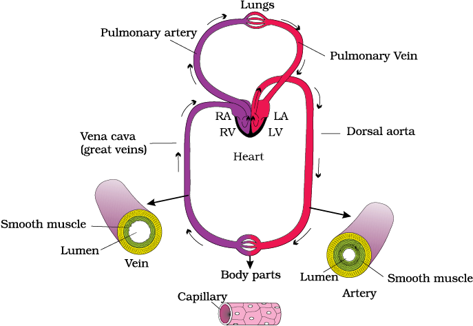
Figure 18.4 Schematic plan of blood circulation in human
© 2024 GoodEd Technologies Pvt. Ltd.