Mechanism of muscle contraction is best explained by the sliding filament theory which states that contraction of a muscle fibre takes place by the sliding of the thin filaments over the thick filaments.
Muscle contraction is initiated by a signal sent by the central nervous system (CNS) via a motor neuron. A motor neuron alongwith the muscle fibres connected to it constitute a motor unit. The junction between a motor neuron and the sarcolemma of the muscle fibre is called the neuromuscular junction or motor-end plate. A neural signal reaching this junction releases a neurotransmitter (Acetyl choline) which generates an action potential in the sarcolemma.
Figure 20.4Stages in cross bridge formation, rotation of head and breaking of cross bridge
This spreads through the muscle fibre and causes the release of calcium ions into the sarcoplasm. Increase in Ca++ level leads to the binding of calcium with a subunit of troponin on actin filaments and thereby remove the masking of active sites for myosin. Utilising the energy from ATP hydrolysis, the myosin head now binds to the exposed active sites on actin to form a cross bridge (Figure 20.4). This pulls the attached actin filaments towards the centre of ‘A’ band. The ‘Z’ line attached to these actins are also pulled inwards thereby causing a shortening of the sarcomere, i.e., contraction. It is clear from the above steps, that during shortening of the muscle, i.e., contraction, the ‘I’ bands get reduced, whereas the ‘A’ bands retain the length (Figure 20.5). The myosin, releasing the ADP and P1 goes back to its relaxed state. A new ATP binds and the cross-bridge is broken (Figure 20.4). The ATP is again hydrolysed by the myosin head and the cycle of cross bridge formation and breakage is repeated causing further sliding. The process continues till the Ca++ ions are pumped back to the sarcoplasmic cisternae resulting in the masking of actin filaments. This causes the return of ‘Z’ lines back to their original position, i.e., relaxation. The reaction time of the fibres can vary in different muscles. Repeated activation of the muscles can lead to the accumulation of lactic acid due to anaerobic breakdown of glycogen in them, causing fatigue. Muscle contains a red coloured oxygen storing pigment called myoglobin. Myoglobin content is high in some of the muscles which gives a reddish appearance. Such muscles are called the Red fibres. These muscles also contain plenty of mitochondria which can utilise the large amount of oxygen stored in them for ATP production. These muscles, therefore, can also be called aerobic muscles. On the other hand, some of the muscles possess very less quantity of myoglobin and therefore, appear pale or whitish. These are the White fibres. Number of mitochondria are also few in them, but the amount of sarcoplasmic reticulum is high. They depend on anaerobic process for energy.
Figure 20.5Sliding-filament theory of muscle contraction (movement of the thin filaments and the relative size of the I band and H zones)
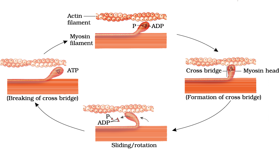 Figure 20.4 Stages in cross bridge formation, rotation of head and breaking of cross bridge

