Skeletal system consists of a framework of bones and a few cartilages. This system has a significant role in movement shown by the body. Imagine chewing food without jaw bones and walking around without the limb bones. Bone and cartilage are specialised connective tissues. The former has a very hard matrix due to calcium salts in it and the latter has slightly pliable matrix due to chondroitin salts. In human beings, this system is made up of 206 bones and a few cartilages. It is grouped into two principal divisions – the axial and the appendicular skeleton.
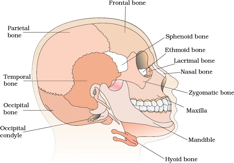
Axial skeleton comprises 80 bones distributed along the main axis of the body. The skull, vertebral column, sternum and ribs constitute axial skeleton. The skull (Figure 20.6) is composed of two sets of bones – cranial and facial, that totals to 22 bones. Cranial bones are 8 in number. They form the hard protective outer covering, cranium for the brain. The facial region is made up of 14 skeletal elements which form the front part of the skull. A single U-shaped bone called hyoid is present at the base of the buccal cavity and it is also included in the skull. Each middle ear contains three tiny bones – Malleus, Incus and Stapes, collectively called Ear Ossicles. The skull region articulates with the superior region of the vertebral column with the help of two occipital condyles (dicondylic skull).
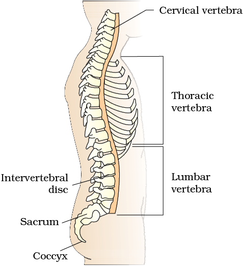
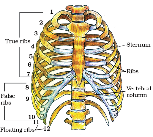
Our vertebral column (Figure 20.7) is formed by 26 serially arranged units called vertebrae and is dorsally placed. It extends from the base of the skull and constitutes the main framework of the trunk. Each vertebra has a central hollow portion (neural canal) through which the spinal cord passes. First vertebra is the atlas and it articulates with the occipital condyles. The vertebral column is differentiated into cervical (7), thoracic (12), lumbar (5), sacral (1-fused) and coccygeal (1-fused) regions starting from the skull. The number of cervical vertebrae are seven in almost all mammals including human beings. The vertebral column protects the spinal cord, supports the head and serves as the point of attachment for the ribs and musculature of the back. Sternum is a flat bone on the ventral midline of thorax.
The bones of the limbs alongwith their girdles constitute the appendicular skeleton. Each limb is made of 30 bones. The bones of the hand (fore limb) are humerus, radius and ulna, carpals (wrist bones – 8 in number), metacarpals (palm bones – 5 in number) and phalanges (digits – 14 in number) (Figure 20.9). Femur (thigh bone – the longest bone), tibia and fibula, tarsals (ankle bones – 7 in number), metatarsals (5 in number) and phalanges (digits – 14 in number) are the bones of the legs (hind limb) (Figure 20.10). A cup shaped bone called patella cover the knee ventrally (knee cap).
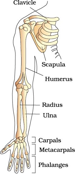
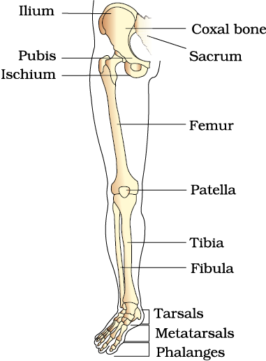
Figure 20.10 Right pelvic girdle and lower limb bones (frontal view)
Pectoral and Pelvic girdle bones help in the articulation of the upper and the lower limbs respectively with the axial skeleton. Each girdle is formed of two halves. Each half of pectoral girdle consists of a clavicle and a scapula (Figure 20.9). Scapula is a large triangular flat bone situated in the dorsal part of the thorax between the second and the seventh ribs. The dorsal, flat, triangular body of scapula has a slightly elevated ridge called the spine which projects as a flat, expanded process called the acromion. The clavicle articulates with this. Below the acromion is a depression called the glenoid cavity which articulates with the head of the humerus to form the shoulder joint. Each clavicle is a long slender bone with two curvatures. This bone is commonly called the collar bone.
Pelvic girdle consists of two coxal bones (Figure 20.10). Each coxal bone is formed by the fusion of three bones – ilium, ischium and pubis. At the point of fusion of the above bones is a cavity called acetabulum to which the thigh bone articulates. The two halves of the pelvic girdle meet ventrally to form the pubic symphysis containing fibrous cartilage.
© 2026 GoodEd Technologies Pvt. Ltd.

