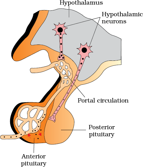
The pituitary gland is located in a bony cavity called sella tursica and is attached to hypothalamus by a stalk (Figure 22.2). It is divided anatomically into an adenohypophysis and a neurohypophysis. Adenohypophysis consists of two portions, pars distalis and pars intermedia. The pars distalis region of pituitary, commonly called anterior pituitary, produces growth hormone (GH), prolactin (PRL), thyroid stimulating hormone (TSH), adrenocorticotrophic hormone (ACTH), luteinizing hormone (LH) and follicle stimulating hormone (FSH). Pars intermedia secretes only one hormone called melanocyte stimulating hormone (MSH). However, in humans, the pars intermedia is almost merged with pars distalis. Neurohypophysis (pars nervosa) also known as posterior pituitary, stores and releases two hormones called oxytocin and vasopressin, which are actually synthesised by the hypothalamus and are transported axonally to neurohypophysis.

Figure 22.2 Diagrammatic representation of pituitary and its relationship with hypothalamus
Over-secretion of GH stimulates abnormal growth of the body leading to gigantism and low secretion of GH results in stunted growth resulting in pituitary dwarfism. Excess secretion of growth hormone in adults especially in middle age can result in severe disfigurement (especially of the face) called Acromegaly, which may lead to serious complications, and premature death if unchecked. The disease is hard to diagnose in the early stages and often goes undetected for many years, until changes in external features become noticeable. Prolactin regulates the growth of the mammary glands and formation of milk in them. TSH stimulates the synthesis and secretion of thyroid hormones from the thyroid gland. ACTH stimulates the synthesis and secretion of steroid hormones called glucocorticoids from the adrenal cortex. LH and FSH stimulate gonadal activity and hence are called gonadotrophins. In males, LH stimulates the synthesis and secretion of hormones called androgens from testis. In males, FSH and androgens regulate spermatogenesis. In females, LH induces ovulation of fully mature follicles (graafian follicles) and maintains the corpus luteum, formed from the remnants of the graafian follicles after ovulation. FSH stimulates growth and development of the ovarian follicles in females. MSH acts on the melanocytes (melanin containing cells) and regulates pigmentation of the skin. Oxytocin acts on the smooth muscles of our body and stimulates their contraction. In females, it stimulates a vigorous contraction of uterus at the time of child birth, and milk ejection from the mammary gland. Vasopressin acts mainly at the kidney and stimulates resorption of water and electrolytes by the distal tubules and thereby reduces loss of water through urine (diuresis). Hence, it is also called as anti-diuretic hormone (ADH).
An impairment affecting synthesis or release of ADH results in a diminished ability of the kidney to conserve water leading to water loss and dehydration. This condition is known as Diabetes Insipidus.

