Following double fertilisation, events of endosperm and embryo development, maturation of ovule(s) into seed(s) and ovary into fruit, are collectively termed post-fertilisation events.
Endosperm development precedes embryo development. Why? The primary endosperm cell divides repeatedly and forms a triploid endosperm tissue. The cells of this tissue are filled with reserve food materials and are used for the nutrition of the developing embryo. In the most common type of endosperm development, the PEN undergoes successive nuclear divisions to give rise to free nuclei. This stage of endosperm development is called free-nuclear endosperm. Subsequently cell wall formation occurs and the endosperm becomes cellular. The number of free nuclei formed before cellularisation varies greatly. The coconut water from tender coconut that you are familiar with, is nothing but free-nuclear endosperm (made up of thousands of nuclei) and the surrounding white kernel is the cellular endosperm.
Endosperm may either be completely consumed by the developing embryo (e.g., pea, groundnut, beans) before seed maturation or it may persist in the mature seed (e.g. castor and coconut) and be used up during seed germination. Split open some seeds of castor, peas, beans, groundnut, fruit of coconut and look for the endosperm in each case. Find out whether the endosperm is persistent in cereals – wheat, rice and maize.
Embryo develops at the micropylar end of the embryo sac where the zygote is situated. Most zygotes divide only after certain amount of endosperm is formed. This is an adaptation to provide assured nutrition to the developing embryo. Though the seeds differ greatly, the early stages of embryo development (embryogeny) are similar in both monocotyledons and dicotyledons. Figure 2.13 depicts the stages of embryogeny in a dicotyledonous embryo. The zygote gives rise to the proembryo and subsequently to the globular, heart-shaped and mature embryo.
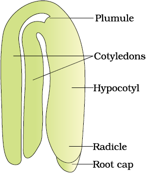
A typical dicotyledonous embryo (Figure 2.14a), consists of an embryonal axis and two cotyledons. The portion of embryonal axis above the level of cotyledons is the epicotyl, which terminates with the plumule or stem tip. The cylindrical portion below the level of cotyledons is hypocotyl that terminates at its lower end in the radicle or root tip. The root tip is covered with a root cap.
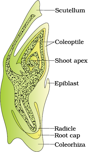
Embryos of monocotyledons (Figure 2.14 b) possess only one cotyledon. In the grass family the cotyledon is called scutellum that is situated towards one side (lateral) of the embryonal axis. At its lower end, the embryonal axis has the radical and root cap enclosed in an undifferentiated sheath called coleorrhiza. The portion of the embryonal axis above the level of attachment of scutellum is the epicotyl. Epicotyl has a shoot apex and a few leaf primordia enclosed in a hollow foliar structure, the coleoptile.
Soak a few seeds in water (say of wheat, maize, peas, chickpeas, ground nut) overnight. Then split the seeds and observe the various parts of the embryo and the seed.
© 2026 GoodEd Technologies Pvt. Ltd.

