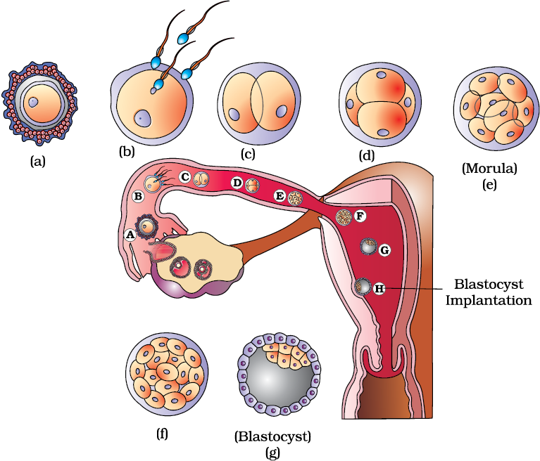
During copulation (coitus) semen is released by the penis into the vagina (insemination). The motile sperms swim rapidly, pass through the cervix, enter into the uterus and finally reach the ampullary region of the fallopian tube (Figure 3.11b). The ovum released by the ovary is also transported to the ampullary region where fertilisation takes place. Fertilisation can only occur if the ovum and sperms are transported simultaneously to the ampullary region. This is the reason why not all copulations lead to fertilisation and pregnancy.
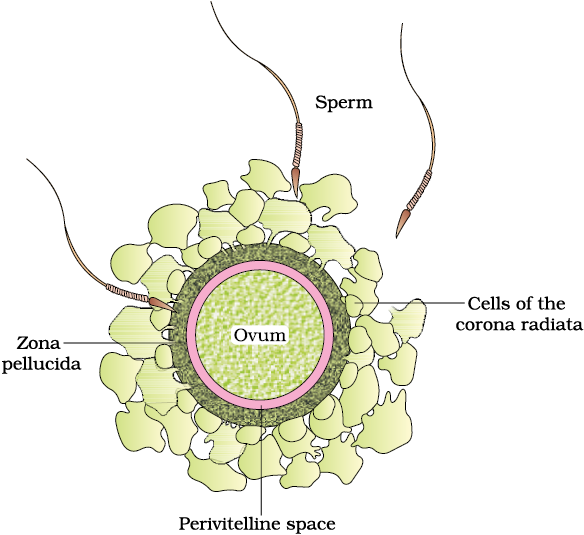
The process of fusion of a sperm with an ovum is called fertilisation. During fertilisation, a sperm comes in contact with the zona pellucida layer of the ovum (Figure 3.10) and induces changes in the membrane that block the entry of additional sperms. Thus, it ensures that only one sperm can fertilise an ovum. The secretions of the acrosome help the sperm enter into the cytoplasm of the ovum through the zona pellucida and the plasma membrane. This induces the completion of the meiotic division of the secondary oocyte. The second meiotic division is also unequal and results in the formation of a second polar body and a haploid ovum (ootid). Soon the haploid nucleus of the sperms and that of the ovum fuse together to form a diploid zygote. How many chromosomes will be there in the zygote?
Figure 3.10 Ovum surrounded by few sperms

One has to remember that the sex of the baby has been decided at this stage itself. Let us see how? As you know the chromosome pattern in the human female is XX and that in the male is XY. Therefore, all the haploid gametes (ova) produced by the female have the sex chromosome X whereas in the male gametes (sperms) the sex chromosome could be either X or Y, hence, 50 per cent of sperms carry the X chromosome while the other 50 per cent carry the Y. After fusion of the male and female gametes the zygote would carry either XX or XY depending on whether the sperm carrying X or Y fertilised the ovum. The zygote carrying XX would develop into a female baby and XY would form a male (you will learn more about the chromosomal patterns in Chapter 5). That is why, scientifically it is correct to say that the sex of the baby is determined by the father and not by the mother!
The mitotic division starts as the zygote moves through the isthmus of the oviduct called cleavage towards the uterus (Figure 3.11) and forms 2, 4, 8, 16 daughter cells called blastomeres. The embryo with 8 to 16 blastomeres is called a morula (Figure 3.11e). The morula continues to divide and transforms into blastocyst (Figure 3.11g) as it moves further into the uterus. The blastomeres in the blastocyst are arranged into an outer layer called trophoblast and an inner group of cells attached to trophoblast called the inner cell mass. The trophoblast layer then gets attached to the endometrium and the inner cell mass gets differentiated as the embryo. After attachment, the uterine cells divide rapidly and covers the blastocyst. As a result, the blastocyst becomes embedded in the endometrium of the uterus (Figure 3.11h). This is called implantation and it leads to pregnancy.
