
 Q. No.
The right atria of the human heart receive:
1. Oxygenated blood
2. Deoxygenated blood
3. Arterial blood
4. Venous blood
1. Oxygenated blood
2. Deoxygenated blood
3. Arterial blood
4. Venous blood


The following is the diagrammatic representation of standard ECG. Identify the correct statement :
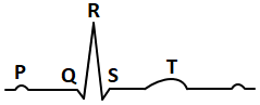
| 1. | P is caused by atrial repolarization. |
| 2. | Q, when present, always shows myocardial ischemia. |
| 3. | QRS complex is due to time taken by impulse from SA node to AV node. |
| 4. | T is caused by ventricular repolarization. |
In the given section of the human heart which part ensures that atria contact prior to the ventricles ?

1. A
2. B
3. C
4. D


Name the blood cells, whose reduction in number can cause clotting disorder, leading to excessive loss of blood from the body.
(1) Erythrocytes
(2) Leucocytes
(3) Neutrophils
(4) Thrombocytes
Serum differs from blood in
(1) lacking globulins
(2) lacking albumins
(3) lacking clotting factors
(4) lacking antibodies
Doctors use a stethoscope to hear the sounds produced during each cardiac cycle. The
second sound is heard when
1. AV valves open up
2. Ventricular walls vibrate due to gushing in of blood from atria
3. Semilunar valves close down after the blood flows into vessels from ventricles
4. AV node receives the signal from SA node
Person with blood group AB is considered as universal recipient because he has
1. Both A and B antigens on RBC but no antibodies in the plasma
2. Both A and B antibodies in the plasma
3. No antigen on RBC and antibody in the plasma
4. Both A and B antigens in the plasma but no antibodies.
How do parasympathetic neural signals affect the working of the heart?
1. Reduce both heart rate and cardiac output
2. heart rate is increased without affecting the cardiac output
3. Both heart rate and cardiac output increase
4. Heart rate decreases but cardiac output increases
The diagram given here is the standard ECG of a normal person. the P-wave represents
the
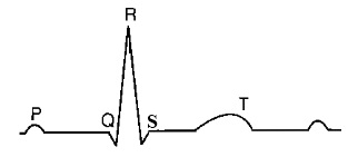
1. contraction of both the atria
2. initiation of the ventricular contraction
3. beginning of the systole
4. end of systole
© 2024 GoodEd Technologies Pvt. Ltd.